Evanratzan.com

 Postdoctoral Research

I currently work as a Postdoctoral Research Fellow in the lab of Jeffrey Holt and Gwenaelle Geleoc at Boston Children’s Hospital Department of Otolaryngology/Harvard Medical School Department of Neurology. The main focus of my research efforts are utilizing gene therapy to restore loss of function in mouse models of congenital sensory loss. Mutations in several different genes (Tmc1, Ush1c, Cib2, Tmie) in mouse models recapitulates human phenotypes. Specifically, the loss of hearing, balance, and in the case of Usher Syndrome vision as well. We utilize adeno associated viruses (AAVs), lipid nanoparticles, and antisense oligonucleotides as a means for addressing underlying genetic causes of sensory loss.

Additionally, many genes that involve loss of function are key components of the mechanoelectrical transduction channel (MET) complex found in hair cells of the inner ear. I am interested in the contribution of these components to the function of organs of the inner ear during development of behaviors. Unlike classic voltage-gated ion channels, the assembly of the MET complex requires several protein components to confer normal mechanosensitivity (TMC1, TMIE, CIB2, and LHFLP5 among others). The expression of these proteins may confer unique characteristics to hair cell subtypes during their development, which may have implicaitons for their function and response to gene therapy.


PhD Research

Untitled-1.gif

Polarized Patterning of the Vestibular Maculae

The utricle and saccule (vestibular maculae) are otolith organs of the inner ear that contain sensory cells that are essential for inner ear function. Among these cells are mechanotransductive hair cells which detect deflection in a specific vector via their apically localized stereociliary bundles. A single tubulin rich kinocilium sits alongside the actin-dense bundles where it is positioned asymmetrically indicative of the maximum deflection sensitivity of a single hair cell. Hair cells are arranged in the utricle and saccule in opposing orientations about a line of polarity reversal (LPR). The formation of this LPR depends on a transcription factor Emx2. But the protein effectors downstream of Emx2 which move the kinocilium to its asymmetric position, had not yet been identified. The current goal of my PhD dissertation was to identify what effector proteins give rise to this polarized pattern. I successfully identified that serine threonine kinase 32 a (Stk32a) was expressed on the opposite side of the LPR as Emx2. Emx2 represses Stk32a expression in hair cells in one domain causing a reversal of hair cell orientation.


20201009-Cover Image3.jpg

During development the vestibular sensory end organs generate both Type-I and Type-II hair cells, however these two classes of sensory cells cannot be distinguished until later postnatal stages when they become morphologically and physiologically distinct. The molecular signaling pathways that establish vestibular hair cell identity have not been clearly defined but are of utmost importance due to the capacity for these cells to regenerate following ototoxic damage. Presumably, regeneration-based therapies will need to generate the correct ratio of hair cell types for the overall physiological responses to be effective.  As a result, understanding the developmental mechanisms contributing to the specification and differentiation of Type-I versus Type-II hair cells has clinical significance. We have found that Fgf8 is expressed in a subset of vestibular hair cells and that the transgenic line FGF8-CreER genetically labels Type-I hair cells specifically, and at the earliest stages of their development. You can read the full story in our published work here.

 
cochlea2.jpeg

Spiral Ganglion Neuron Turning in the Developing Cochlea

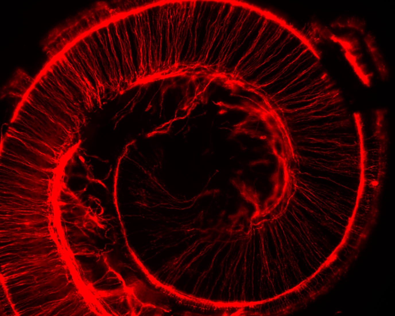
The cochlea is a snail-shell shaped organ within our inner ear that also contains hair cells which meachanically transduce pressure waves into meaningful sounds for our brain. These hair cells are innervated by spiral ganglion neurons (SGNs) which are so named because of their characteristic spiraling turns from base to apex along the cochlear length. Recent work by my fellow labmate Satish Ghimire revealed that the transmembrane protein Van Gogh Like 2 (Vangl2) plays an important role in this process. Vangl2 is known to have a cell-autonomous effect in axon guidance in the spinal cord. However, uniquely we found that there was a non-autonomous function of Vangl2 from the nearby environment which has an effect on the correct guidance and innervation of hair cells in the cochlea by type II SGNs. To learn more about this research, please read our publication a non-autonomous function of the core PCP protein VANGL2 directs peripheral axon turning in the developing cochlea.


Optogenetics.JPG

The Effect of Stress on Drug Seeking Behavior

For two years I worked with Dr. Michael Baratta, PhD at the University of Colorado Institute of Behavioral Genetics investigating the role of stress in mouse and rat models of cocaine and nicotine addiction. We used a combination of optogenetics and designer receptor exclusively activated by designer drugs (DREADDS) in order to manipulate the perforant path. We observed how different stressors influenced rat drug seeking behavior, and examined the role of coping mechanisms in attenuating addiction. My primary responsibilities included construction of optogenetic implants, fiber optics, behavioral training apparatuses, and craniotomy/viral injections of experimental animals.

The Role of Glutathione in Mercury Clearance from Human Blood

Quicksilver Scientific was founded by Christopher Shade, PhD, N.D. who developed a novel method of extracting different species of mercury from a variety of matrices including environmental and clinical samples. Since its emergence, Quicksilver has expanded clinical operations substantially and developed unique chelation products and liposomal delivery systems for detoxification. During my employment, my primary goal was to develop a protocol to assess levels of glutathione, glutathione transferase, and glutathione peroxidase from human blood and correlate these levels with levels of inorganic and methyl mercury in the same subjects. Glutathione is a known detoxification agent containing sulfur moieties which likely bind strongly to mercury, and we hypothesized that it played a role in removing mercury contaminants from subjects. I spent additional time in the lab aiding in the synthesis of intestinal metal detox chelator (IMD), and extracting mercury for HPLC analysis from clinical (blood, hair, urine) and environmental samples (soil, water, fish).

Separation, Isolation, & Purification of Transition Metals and Rare Earth Elements

Tusaar Corp. originally emerged from technology developed by the University of Colorado Department of Environmental Engineering designed to extract metals from aqueous streams. Since its inception, Tusaar has expanded and built upon this technology to also include toxic metal sequestration, precious metal recovery, and separation/purification of metals. My primary responsibilities as an Applications Engineer included bench level testing of client samples, construction/implementation of scaled-up onsite pilot studies, and quality control of outgoing material. Initially, these projects targeted removal of toxic and heavy metals from aqueous waste streams and concentrating them into a non-leachable solid media for disposal. However, projects later emerged in which we would target specific rare earth elements from recycled or abandoned solid wastes for extraction and purification.